

October 26, 2026 · 12 min read
Shoulder Injuries: An Evaluation and Referral Framework for Primary Care Physicians
Medically reviewed content from RegenOrthoSport, Dallas, Texas.
Orient the clinical conversation around assessment, not labels
Shoulder injury assessment works best when you connect the history, functional effect, examination, and clinical question before choosing a label. Shoulder pain can arise from joints, tendons, nerves, blood vessels, bones, or nearby structures, so the same complaint can lead to different evaluation paths. The cited acute-shoulder guidance defines acute pain as lasting less than six months. The cited chronic-shoulder guidance generally classifies pain lasting more than six months as chronic. Those time frames organize the conversation; they do not identify the cause.
Your first task is to clarify what happened and what has changed. Ask how the problem began, which movements or activities aggravate it, whether weakness or tingling accompanies the pain, and how the problem affects work, recreation, or daily movement. Then use the examination to decide whether the concern fits a routine conservative pathway, needs imaging, or merits specialist assessment.
The framework below is designed for patient counseling and referral decisions. It does not diagnose a specific injury or determine eligibility for a particular treatment. Use it to make the next clinical question more precise.
Map the structures commonly involved in shoulder injuries
Shoulder injuries can affect several connected structures, and the involved structure changes what you need to examine. Many shoulder problems involve muscles, ligaments, or tendons, but fractures can affect the humerus or glenoid. Trauma can also involve the clavicle, scapula, joints, neurovascular structures, or nearby anatomy.
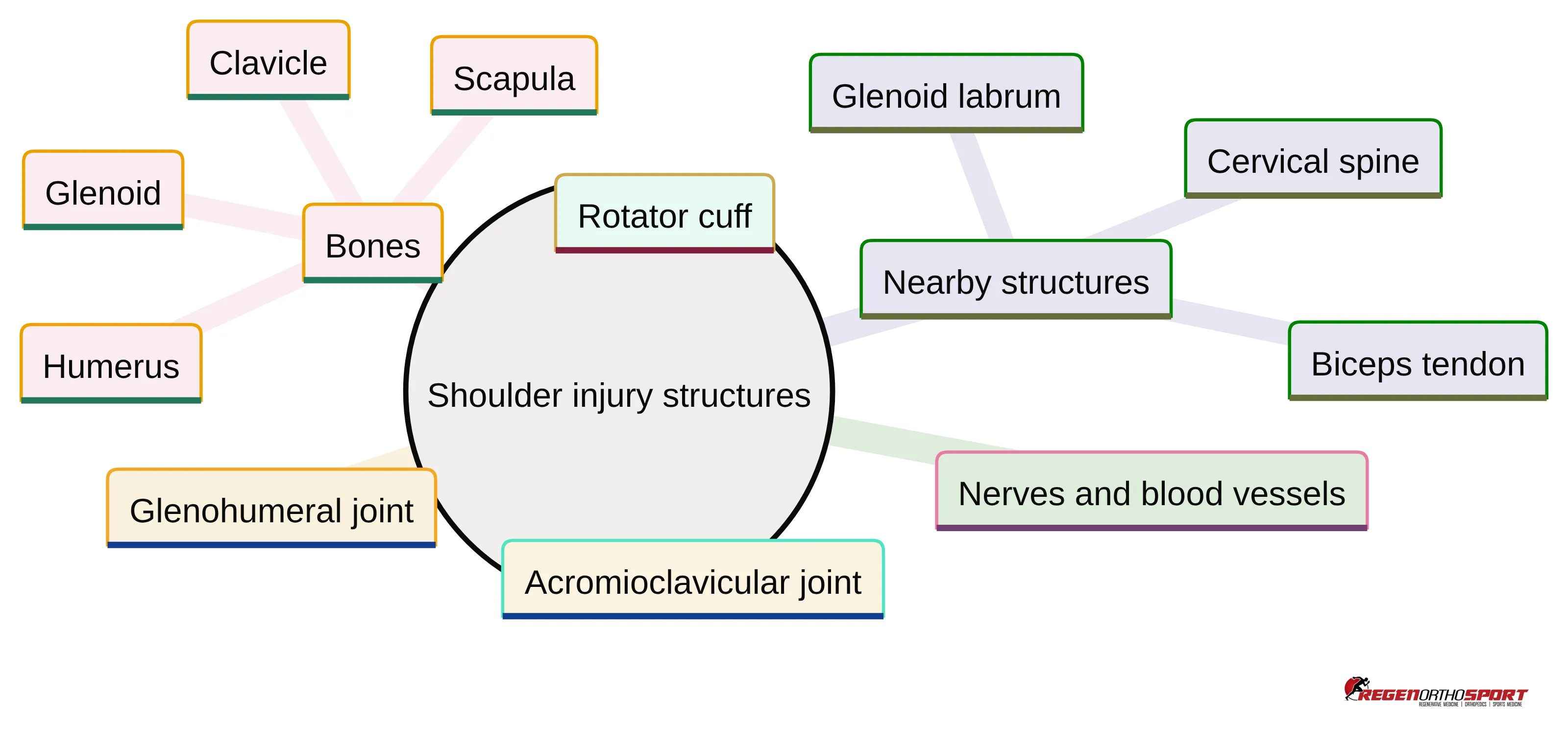
Use the following map to keep the history broad enough at the start:
- Rotator cuff: Tendon injuries include tendinosis and partial or full-thickness tears. Rotator cuff problems are common in primary care and may require soft-tissue imaging when the clinical question remains after the initial assessment.
- Acromioclavicular joint: Problems in this joint may follow a fall or direct impact onto the shoulder. Acromioclavicular joint disease is among the common shoulder problems seen in primary care.
- Glenohumeral joint: This joint may be involved in joint disease, instability, or dislocation-related injury. History and examination help organize the concern before specialist imaging is considered.
- Bones: The humerus, clavicle, scapula, and glenoid can be affected by trauma. A separated shoulder after a fall and fractures after high-energy trauma represent different injury contexts.
- Nerves and blood vessels: Neurovascular structures deserve attention when weakness, tingling, or other sensory changes accompany shoulder symptoms.
- Nearby structures: The glenoid labrum, biceps tendon, and cervical spine can contribute to symptoms described as shoulder pain.
This map prevents the conversation from narrowing prematurely to the rotator cuff. Keep the structure under consideration aligned with the mechanism, examination, and functional change.
Describe symptoms through their effect on function
Shoulder symptoms become more useful clinically when you describe what they prevent or change. Pain with overhead elevation, weakness during the same movement, and tingling are different findings that can point the history in different directions, but none establishes a diagnosis by itself.
Ask the patient to describe function in concrete terms:
- Which movement brings on the symptom: overhead elevation, reaching across the body, lifting, or another activity?
- Does the limitation come from pain, loss of strength, restricted motion, or a combination?
- Has the problem changed work, exercise, domestic activity, or leisure activity?
- Does the symptom appear with a particular task, or does it persist outside that task?
- Is tingling present, and does it occur with weakness or without it?
Pain and weakness during overhead elevation can occur with a rotator cuff tear or suprascapular nerve injury. The finding is therefore a reason to broaden the assessment, not a diagnostic rule. When pain, weakness, or tingling occurs without a known bone, soft-tissue, or vascular injury, possible upper-extremity nerve injury belongs in the differential.
Functional restriction also helps you explain why assessment matters. A patient may tolerate a symptom at rest yet have a substantial restriction during work or sport. Record the activity and the specific limitation rather than reducing the encounter to a pain score alone.
Use mechanism and time course to organize the history
Shoulder injury history starts with mechanism and time course because a fall, direct impact, repetitive overhead activity, and gradual onset create different assessment questions. Acute shoulder injuries commonly follow a fall or direct trauma in contact and collision sports. A symptom that develops gradually during a repeated activity can require a different line of questioning.
A focused history can follow this sequence:
- Reconstruct the event. Ask whether the problem followed a fall, direct blow, collision, lifting event, or another activity. Clarify the position of the arm and whether the shoulder was forced beyond its usual movement.
- Establish the timeline. Record the date of onset, whether symptoms are improving or persisting, and whether the problem has lasted less than or more than six months. The acute and chronic time frames help organize evaluation but do not name the injury.
- Connect symptoms to activity. Ask what movement aggravates the shoulder and whether the timing is consistent. Upper-extremity nerve symptoms can be acute or gradual, and the relationship between activity and symptom onset can be clinically useful.
- Identify functional change. Document the task the patient cannot perform, performs differently, or avoids because of pain, weakness, restricted motion, or tingling.
- Ask about associated findings. Include weakness, paresthesias, and the location of symptoms, while keeping referred pain from the cervical spine in consideration.
The history should narrow the examination question without turning a symptom pattern into a diagnosis. Move to examination when the mechanism, duration, and functional effect are clear enough to guide that next step.
Make the examination the bridge to the next decision
Shoulder examination connects the patient’s story to the next decision. Shoulder instability is assessed through history and examination.
An initial examination should assess and compare the injured and uninjured sides:
- Bones: Examine the humerus, clavicle, scapula, and areas of focal tenderness or altered contour.
- Joints: Consider the acromioclavicular, sternoclavicular, and glenohumeral regions in relation to the mechanism and movement limitation.
- Soft tissues: Assess the rotator cuff region and other involved soft tissues in the context of pain, weakness, and motion.
- Neurovascular structures: Check findings that could support concern for nerve or blood-vessel involvement, especially when tingling or weakness is present.
- Function: Compare active movement and the task that the patient identifies as limited.
Your documentation should show how the examination supports the next question. For example, persistent overhead weakness may justify a focused assessment of the rotator cuff and possible nerve involvement, while trauma with focal bony findings may shift attention toward radiography. These patterns guide evaluation; they do not establish a diagnosis without the full clinical assessment.
If the mechanism, examination, or functional change does not fit a routine explanation, keep the assessment open and consider specialist input rather than offering reassurance based on a single finding.
Match imaging to the clinical question and symptom duration
Shoulder imaging should answer a clinical question that follows the history and examination. After history and examination, plain radiography is commonly used as the initial imaging study for acute shoulder injuries. For chronic shoulder pain, the cited ACR criteria rate shoulder radiography as usually appropriate for initial imaging.
The distinction matters because radiography and soft-tissue imaging show different structures. X-rays do not show the rotator cuff and may be normal with rotator cuff pain. MRI or ultrasound can visualize the tendons and characterize a tear. A normal radiograph therefore does not settle a rotator cuff question.
Use the symptom duration and suspected structure to organize the next step:
- Acute injury: Begin with the mechanism, examination, and plain radiography when the clinical assessment calls for imaging.
- Chronic shoulder pain: The cited ACR criteria rate shoulder radiography as usually appropriate as the initial study.
- Suspected rotator cuff disorder or subacromial bursitis after normal or inconclusive radiographs: The cited ACR criteria rate shoulder ultrasound as usually appropriate as the next imaging study.
- Soft-tissue characterization: MRI or ultrasound can show the rotator cuff tendons, the location of a tear, and its size; the choice depends on the clinical question and assessment pathway.
Imaging should add information to the examination rather than replace it. Explain to the patient what the selected study is intended to clarify, and avoid treating an imaging finding as the whole diagnosis without matching it to symptoms and function.
Recognize when the available information is not enough for routine reassurance
Shoulder injury information is not sufficient for routine reassurance when the mechanism, functional loss, or neurologic finding raises a separate assessment question. Your role is not to force every presentation into a common label; your role is to recognize when the next step needs more evaluation.
Arrange clinical assessment rather than relying on reassurance alone when you identify circumstances such as:
- A fall, direct impact, collision, or high-energy event with focal shoulder findings or marked functional change.
- New weakness or tingling without a known bone, soft-tissue, or vascular injury, because possible upper-extremity nerve injury can mimic common musculoskeletal disorders.
- Shoulder pain and weakness during overhead elevation when the findings could reflect either rotator cuff or suprascapular nerve involvement.
- Persistent symptoms lasting more than six months, particularly when work, domestic activity, or leisure remains restricted.
- Examination findings that do not match the reported mechanism or that cannot be localized confidently.
Shoulder trauma can include separation after a fall or fractures of the scapula or clavicle after high-energy trauma. The mechanism alone does not determine the injury, but it should affect the threshold for a careful examination and appropriate imaging. Similarly, tingling or weakness should not be explained away as ordinary shoulder pain without considering nerve involvement.
Use the uncertainty itself as a reason to clarify the assessment. A patient does not need a premature label before you decide that further evaluation or referral is appropriate. Duration should be interpreted with functional limitation, worsening symptoms, concerning examination findings, or diagnostic uncertainty rather than treated as a referral threshold by itself.
Start with condition-appropriate non-surgical care
Non-surgical care should match the condition’s phase, symptom severity, functional restrictions, and response to care. The cited guidance states that most acute shoulder injuries can be managed conservatively with comfort measures and targeted rehabilitation, while some injury patterns require specialist assessment.
For a patient whose assessment supports a conservative pathway, discuss the categories that may be considered:
- Comfort measures and activity modification: A sling may be used for comfort in selected acute injuries, and limiting activities that aggravate the shoulder may be considered.
- Medication discussion: Medicines such as NSAIDs may be part of care when clinically appropriate for the patient. Selection and use require attention to the patient’s medical context.
- Targeted rehabilitation: A clinician-guided program may address flexibility, range of motion, and strengthening. The exercises should be selected with a physician or physical therapist rather than copied from a generic routine.
- Follow-up based on function: Reassess symptoms, movement, strength, and the patient’s ability to perform relevant work, domestic, or leisure activities.
Conservative rotator cuff care may include activity modification, NSAIDs, and clinician-guided flexibility, strengthening, or physical therapy. The purpose of the plan is not to assume the diagnosis; it is to provide a condition-appropriate pathway while the clinical picture is monitored.
Treatment should change when the examination, symptom course, or functional restriction changes. Explain that non-surgical treatment selection requires communication with a healthcare provider and following the recommended treatment plan. Avoid promising a particular result, and do not select rehabilitation or medication solely from the word rotator cuff.
Place PRP therapy and stem cell therapy within individualized evaluation
PRP therapy and stem cell therapy should be considered only after the injury pattern, examination, symptom duration, functional restriction, imaging question, and response to initial care have been reviewed. This overview does not establish an indication, effectiveness, or safety profile for either option; individual experiences with shoulder pain treatment may vary. The injury pattern, examination, symptom duration, functional restriction, imaging question, and response to initial care should be clear enough to support a meaningful discussion before any option is selected.
Use a structured conversation with the patient:
- Name the clinical question first. Clarify whether the concern involves the rotator cuff, a joint, trauma-related structures, a nerve, or another area.
- Separate evaluation from treatment choice. A treatment label cannot replace an examination or determine whether the option fits the injury.
- Address uncertainty directly. Individual experiences with shoulder pain treatment may vary.
- Document the decision basis. Record the assessment, relevant findings, functional effect, prior care, and the reason for referral or treatment discussion.
Our stem cell therapy process involves collecting and concentrating bone marrow, then injecting it into an injured or arthritic area with ultrasound or X-ray guidance. That description explains our process; it does not establish that the process fits every shoulder injury or predict an outcome.
A referral conversation can therefore be useful even when the patient is specifically asking about regenerative medicine. Start with the injury context and examination, then discuss whether a consultation about non-surgical options is appropriate. A qualified healthcare professional should determine individual suitability.
Turn the assessment into a Dallas–Fort Worth referral conversation
Dallas–Fort Worth referral decisions become clearer when you send the clinical question with the patient rather than only the symptom label. A useful referral summary can include:
- The mechanism, date of onset, and whether the problem is acute or chronic.
- The movements, work tasks, sports, or daily activities that remain limited.
- Findings from the comparison with the uninjured shoulder, including strength, motion, focal tenderness, and neurovascular observations.
- Imaging already obtained and the question that remains unanswered.
- Conservative care tried, the patient’s response, and the reason further assessment is being considered.
We provide consultations regarding shoulder pain treatment with Dr. Movva at our Dallas office. We offer consultations and procedures at our Dallas office on Walnut Hill Lane and serve patients from across Dallas–Fort Worth. Those are location and service facts, not a conclusion that every shoulder injury requires referral to our practice.
For a patient considering PRP therapy or stem cell therapy, frame the referral around individualized assessment of the shoulder concern and possible non-surgical options. For a patient with uncertain localization, neurologic findings, trauma-related concerns, or persistent functional restriction, send the history and examination details that explain why the clinical question remains open.
A focused referral helps the specialist begin with the relevant context. It also gives you a clearer basis for discussing what is known, what still needs assessment, and which next step fits the patient’s presentation.
Invite discussion before selecting a treatment path
Shoulder injury decisions become more disciplined when you connect mechanism, time course, function, examination, imaging, and response to care. Use the symptom pattern to guide questions, not to make a diagnosis from a single finding. Use imaging to answer a defined clinical question, and place PRP therapy or stem cell therapy after injury-specific assessment rather than before it.
For patients in Dallas–Fort Worth, we provide consultations regarding shoulder pain treatment with Dr. Movva at our Dallas office. Each person’s experience with shoulder pain treatment may vary, so a qualified healthcare professional should determine individual suitability and discuss the appropriate next step.
Give the patient a clear explanation of what you know, what requires further evaluation, and why the proposed referral or treatment discussion fits the current assessment.
Common Questions About Shoulder Injuries and Treatment
What are common symptoms of shoulder injuries?
Common symptoms include pain during overhead movements, weakness, restricted motion, and tingling. These symptoms can indicate various underlying issues, such as rotator cuff tears or nerve injuries.
What initial imaging is typically recommended for acute shoulder injuries?
Plain radiography is usually the initial imaging study for acute shoulder injuries to assess for fractures or other bony abnormalities.
What role do PRP and stem cell therapies play in shoulder injury treatment?
PRP and stem cell therapies are considered after a thorough evaluation and are part of a broader discussion on non-surgical options, tailored to the individual patient’s condition.
What factors should be considered when referring a patient for shoulder injury treatment?
Factors include the mechanism of injury, duration of symptoms, functional limitations, and any previous treatments attempted.